Contact Form

BRAIN ANEURYSMS

What is the brain aneurysm?

A brain aneurysm is a condition characterized by ballooning and expansion of the brain vessel's wall, which can potentially rupture and cause bleeding.

How does the brain get its blood supply?

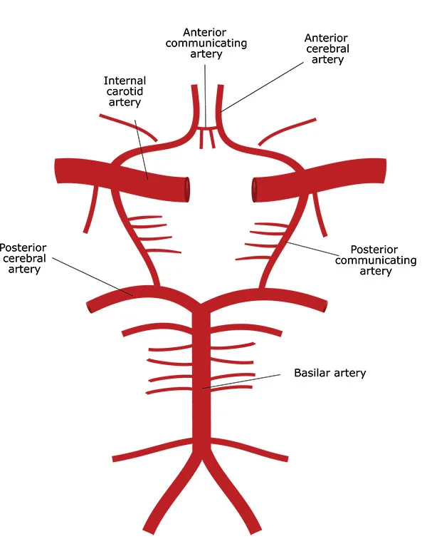
The brain is a very energy-consuming organ. It weighs around 1.5 kg yet takes approximately 15% of total blood flow and 20% of total oxygen consumption. It is supplied by four major vessels - two carotid and two vertebral arteries. These arteries form a complex network called The Circle of Willis, which supplies the brain by giving off smaller branches. The Circle of Willis allows the brain to maintain blood flow in cases of major artery obstruction. It is an evolutional adaptation to keep all brain parts continuously supplied. Technically, brain arteries and circles of Willis are located outside of the brain in the subarachnoid space. This space is filled with cerebrospinal fluid.

Why do the brain arteries develop aneurysms?

Brain Aneurysm Vessels

The flow and the pressure in the brain arteries are very high due to massive demand. Also, the wall of brain arteries lacks an external supporting layer (tunica externa or adventitia layer) present in other arteries. Therefore, brain arteries are very prone to aneurysm formation. The weak spots in the artery wall become bulged due to pressure inside of the artery. These small bulges are sometimes called blister aneurysms because they can also rupture and cause bleeding. With time, this bulge became more and more prominent due to the Laplace law. According to this law, the greater the diameter of the vessel, the greater the tension of its wall, given the pressure is constant. Therefore, bigger aneurysms tend to rupture more frequently.

All brain aneurysms are true aneurysms, unlike false aneurysms that may develop in other areas. Brain aneurysms usually develop at sites where the artery branches off.

Blood flow at branching sites is usually turbulent, putting additional mechanical stress on the arterial wall. Thus, these sites are especially prone to aneurysm formation. High blood pressure (hypertension), smoking, genetic predisposition (familial cases), and atherosclerosis are factors contributing to aneurysm formation.

How many brain aneurysm types are there?

Berry Brain Aneurysm

There are three types of brain aneurysms:

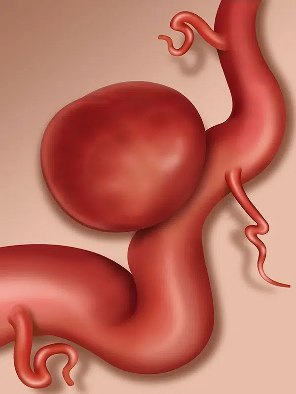
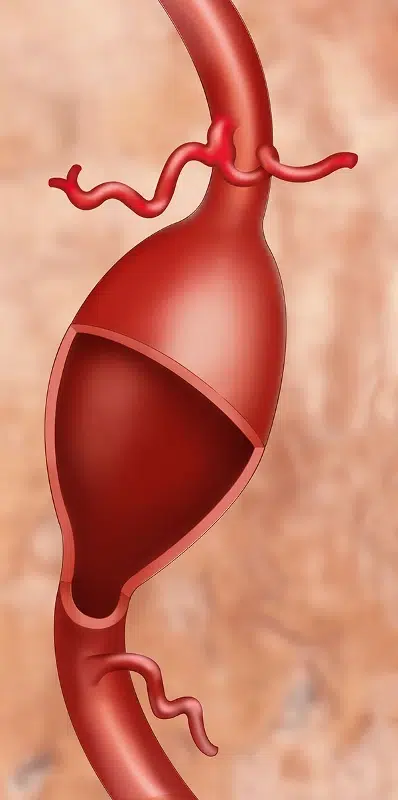
Berry or saccular aneurysms – the most common type of aneurysms. These aneurysms develop due to dilation of the part of the artery wall. They usually have rounded shape and may be from several millimeters to several centimeters.

Usually, these aneurysms become symptomatic due to rupture or compression of nearby structures. They may also develop due to increased flow and turbulence. If one of the main brain arteries is occluded, the others have to compensate through the circle of Willis. Increased flow puts additional mechanical stress on arteries, leading to aneurysm formation.

Brain arteriovenous malformations (AVM) are also frequently associated with aneurysms. AVMs divert blood flow from the brain tissue directly to veins. Since this alternative way has very low resistance, the flow is very high. Therefore, aneurysms often develop on arteries supplying AVM and on the circle of Willis.

Brain Aneurysm Types

Fusiform aneurysms – these aneurysms are dilation of the entire wall of the vessel and resemble a spindle. They also have the potential to rupture, but in the vast majority of cases, they usually cause compression of nearby structures and cause neurological deficits. In some instances, the flow in the aneurysm is slow due to dilation, which leads to blood flow stagnation and thrombus formation. These thrombi may dislodge and cause blood flow obstruction, leading to ischemic stroke.

Mycotic aneurysms – these aneurysms form due to infection. They are usually associated with infective endocarditis. Infected thrombi with bacteria in it may get stuck inside brain vessels. Bacteria will cause inflammation inside the artery wall, leading to weakening and aneurysm formation. These aneurysms usually develop on small brain arteries and are very rare.

What are the symptoms of a brain aneurysm?

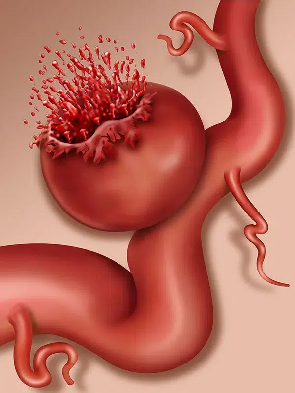
In the vast majority of cases, brain aneurysms do not cause any symptoms until they rupture and cause hemorrhage. Since brain arteries are located in the subarachnoid space, blood spills into this space, causing a subarachnoid hemorrhage.

Ruptured Brain Aneurysm

This type of hemorrhage is associated with the sudden onset of severe headache. Some patients refer to it as the worst pain of their life. This pain is due to irritation of the meninges (brain covers that are very rich with pain receptors).

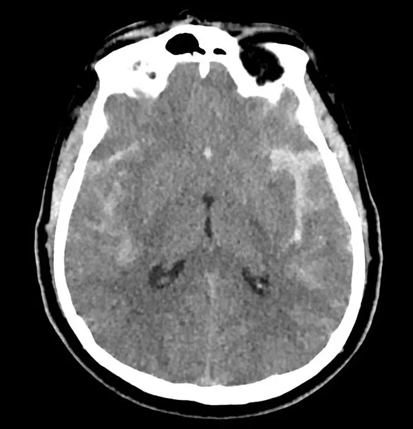
Blood spilled into the subarachnoid space forms clots within minutes after bleeding. Usually, a brain computerized tomography (CT) scan is performed, and bleeding is confirmed. Sometimes, bleeding may be so minuscule that a CT scan may not detect it. Lumbar puncture might be performed on these patients. If bloody cerebrospinal fluid is encountered, the diagnosis is confirmed. Below is a CT scan of a patient with subarachnoid hemorrhage (white areas in the brain).

Brain Aneurysm MRI View

These clots may obstruct cerebrospinal fluid flow and cause acute hydrocephalus. Also, starting several days after hemorrhage, blood clots break down, releasing blood degradation products. This is a very dangerous time period since blood degradation products cause vasospasm - i.e., narrowing of brain arteries. The resultant effect is decreased flow to the brain with ischemic change, further aggravating the patient's condition. In some cases, the hemorrhage may be very massive, and an aneurysm may rupture not only into subarachnoid space but also into the brain itself. Patients may have fallen into a coma and/or develop neurological deficits.

Aneurysms may cause compression of nearby cranial nerves. Second and third cranial nerves are usually very prone to this type of compression, and patients may experience blurry or double vision. Sometimes, aneurysms may be gigantic and cause brain compression, resulting in neurological deficits or epilepsy.

How is a brain aneurysm diagnosed?

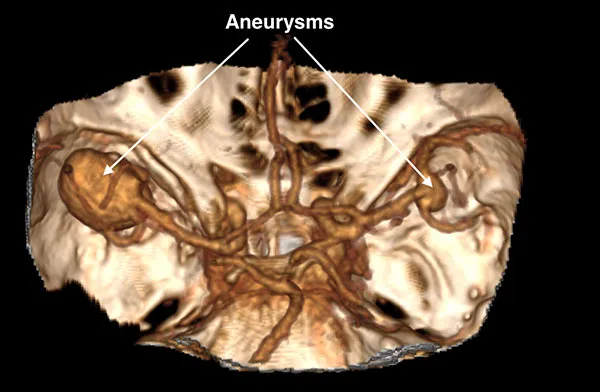
When a brain aneurysm is suspected, brain angiography (literally, vessel imaging) is required to demonstrate its presence, size, and location. Angiography may be performed on CT (CT angiography or CTA), MRI (MR angiography or MRA), or through catheters placed into brain vessels (Digital Subtraction Angiography). Although DSA is considered the gold standard for detecting brain aneurysms, it is an invasive procedure. CTA and MRA nowadays are capable of demonstrating the majority of aneurysms and avoiding DSA procedures. Below is the picture of the CTA of a patient who was referred for spontaneous subarachnoid hemorrhage and was found to have two aneurysms.

Brain Aneurysm Detailed View

What is the treatment for brain aneurysms?

Treatment for aneurysms depends on their rupture status. If the aneurysm is found incidentally on a brain scan, usually, there is no need for urgent intervention. These cases are done electively. However, ruptured aneurysms should be treated as soon as possible to prevent re-bleeding.

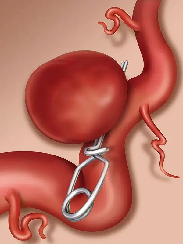
There are two main treatment options for brain aneurysms – microsurgical clipping and endovascular treatment. Microsurgical clipping is performed by neurosurgeons and consists of approaching the aneurysm and placing a permanent clip across the neck of the aneurysm to obstruct the flow into it. This way, an aneurysm is effectively excluded from blood circulation. It is a very effective treatment modality.

The rate of complete occlusion is high, and the recurrence following microsurgical clipping is very low. Another advantage of surgical intervention is the ability to remove blood clots from the brain and decrease the rate of vasospasm and hydrocephalus.

Brain Aneurysm Treatment

Endovascular treatment is a collective term that includes many various types of interventions that aim either to fill the aneurysm or to divert the flow from the aneurysm to reduce the chance of bleeding. These treatments are usually provided either by neurosurgeons or interventional radiologists. The major advantage is avoidance of open brain surgery, yet long-term occlusion rates are not as satisfactory as with microsurgical clipping. The decision to treat a particular aneurysm with surgery or endovascular means should be made by a specific, experienced team practicing both treatment modalities.

Tags: Brain aneurysm symptoms, brain aneurysm diagnosis, brain aneurysm treatment, subarachnoid hemorrhage, cerebral vasospasm, brain aneurysm microsurgical clipping, brain aneurysm endovascular treatment

Aska Question

icon
What are the common symptoms of a brain tumor?
icon

Common symptoms of a brain tumor can vary greatly and depend on the tumor's location, size, and growth rate. They may include headaches, seizures, changes in personality or behavior, memory problems, and difficulty with balance, speech, hearing, or vision. Learn more on this topic in our Brain Tumors section.

Learn More
icon
How is a brain aneurysm diagnosed?
icon

A brain aneurysm is typically diagnosed through imaging methods such as a CT scan, MRI, or cerebral angiography. A lumbar puncture is sometimes done if there is suspicion of an aneurysm rupture. You can read more about this in our dedicated Brain Aneurysms section.

Learn More
icon
What are the current treatment methods for pituitary adenoma?
icon

Treatment options for a pituitary adenoma include observation, medication, surgery, and radiation therapy. The best treatment option depends on the size and type of the tumor, the patient's overall health, and personal preferences. Learn more about Pituitary Adenoma treatment in the corresponding section.

Learn More
icon
What are the causes of disc herniation?
icon

Disc herniation can occur due to aging, physical stress, or injury. As we age, our discs lose some of their water content, increasing the likelihood of tearing or bulging with even slight strain or twist. Learn more about different types of disc herniation in our Spine Diseases section.

Learn More
icon
How is scoliosis treated?
icon

Scoliosis treatment depends on the degree of the curve and the patient's age. Non-surgical treatments include physical therapy, brace use, and pain relievers. In severe cases, surgery may be required to correct the curve. Learn more about scoliosis is treated in our Scoliosis Treatment section.

Learn More
icon
What is degenerative disc disease and what are its main symptoms?
icon

Degenerative disc disease is a condition of wear and tear on the intervertebral discs in the spine with age. Symptoms can include back or neck pain, numbness and tingling sensation, and difficulty in walking. Learn more in our Spine Diseases section.

Learn More
icon
What is thoracic outlet syndrome?
icon

Thoracic outlet syndrome is a condition where the space between the collarbone and the first rib narrows, compressing the nerves or blood vessels. This can lead to pain in the neck and shoulder and numbness in the fingers. Learn more about TOS in our Thoracic Outlet Syndrome section.

Learn More
icon
What are the causes of thoracic outlet syndrome?
icon

Risk factors for thoracic outlet syndrome include poor posture, obesity, repetitive arm or shoulder movements, and anatomical abnormalities such as an abnormal rib or a narrow space between the shoulder and rib. Learn more in our Thoracic Outlet Syndrome section.

Learn More
icon
How is thoracic outlet syndrome diagnosed and treated?
icon

The diagnosis of TOS is made in conjunction with the patient's history, symptoms, physical examination, imaging tests, and neurophysiological tests. Treatment can include physical therapy, pain management, and surgery in severe cases. Learn more about treatment options in our Thoracic Outlet Syndrome Treatment section.

Learn More
icon
What are the signs and symptoms of neurological disorder?
icon

Symptoms of neurological disorders can range widely but commonly include muscle weakness, uncoordinated movements, seizures, persistent or severe headaches, cognitive and memory problems, and changes in sensation. Learn more about neurological disorders in our Brain Diseases section.

Learn More
icon
Are there any warning signs of a brain aneurysm?
icon

Warning signs of a brain aneurysm can encompass sudden and severe headaches, blurred or double vision, neck pain, fainting or dizziness, and sensitivity to light. However, many aneurysms may not show symptoms until they leak or rupture. Learn more about this in our Brain Aneurysms section.

Learn More
icon
What size brain aneurysm requires surgery?
icon

The need for surgery typically depends on the size, location, and growth rate of the aneurysm. Generally, aneurysms larger than 7mm, those located at certain parts of the brain, or those showing signs of growth on consecutive scans may require surgical intervention. To understand the specifics, it's best to consult with a neurosurgeon. More on this topic in our Brain Aneurysms section.

Learn More
icon
Can brain AVM cause personality changes?
icon

Yes, a brain AVM can potentially lead to personality changes if it affects areas of the brain responsible for behavior, cognition, and emotion. Other symptoms might include headaches, seizures, or neurological deficits. Discover more about this disease in our Brain AVM section.

Learn More
icon
Can a blood test detect brain cancer?
icon

Currently, there's no standard blood test that can definitively diagnose brain cancer. While certain markers or changes may suggest a tumor's presence, imaging tests like MRI or CT scans remain primary diagnostic tools. Learn more about the diagnostic processes for brain cancer in our Brain Tumors section.

Learn More
icon
Can an x-ray show a brain tumor?
icon

X-rays are not typically used to detect brain tumors. MRI and CT scans are the primary imaging modalities for this purpose, offering more detailed views of the brain's structure. Explore the specifics of diagnosing brain tumors in our Brain Tumors section.

Learn More
icon
How long does it take for a brain tumor to grow and cause symptoms?
icon

The growth rate of brain tumors can vary based on the type and grade of the tumor. Some tumors grow slowly and might not cause symptoms for years, while others can grow rapidly and present symptoms within weeks or months. The onset of symptoms also depends on the tumor's location and size. Learn more about the progression of brain tumors in our Brain Cancer section.

Learn More
icon
What happens if Chiari malformation goes untreated?
icon

If Chiari malformation goes untreated, symptoms might worsen over time, leading to chronic pain, impaired coordination, and in severe cases, paralysis. Early intervention is recommended to prevent potential complications. Learn more about this in our Chiari Malformation section.

Learn More
icon
Does hemifacial spasm go away?
icon

Hemifacial spasms might diminish with certain treatments, but spontaneous remission is rare. Proper medical intervention can alleviate the symptoms and improve the quality of life. Learn more about the treatment options in our Hemifacial Spasm Treatment section.

Learn More
icon
What happens if hydrocephalus is left untreated?
icon

If left untreated, hydrocephalus can lead to increased intracranial pressure, causing cognitive impairments, vision problems, loss of coordination, and potentially life-threatening brain damage. Learn more about Hydrocephalus Treatment in our dedicated section.

Learn More
icon
What happens if a pituitary tumor goes untreated?
icon

An untreated pituitary tumor can grow and press against essential parts of the brain, disrupting hormonal balances and leading to a variety of health issues, from vision problems to metabolic disorders. Read more about this in our Pituitary Tumors section.

Learn More
icon
What can be mistaken for trigeminal neuralgia?
icon

Trigeminal neuralgia symptoms can be similar to other conditions like dental problems, sinusitis, cluster headaches, or temporomandibular joint disorders, making accurate diagnosis essential. Learn more about how to detect Trigeminal Neuralgia in our dedicated section.

Learn More
icon
What to expect after brain surgery?
icon

After brain surgery, patients might experience fatigue, mood fluctuations, or cognitive changes. Recovery time varies, and regular follow-ups are essential to monitor healing and detect any complications. Learn more about the recovery process for different disorders in our Brain Diseases Treatment section.

Learn More
icon
What happens to the spine as we age?
icon

As we age, the spine undergoes natural degenerative changes. Discs may lose hydration and elasticity, vertebral bones might thin, and there can be a gradual narrowing of the spinal canal, which might lead to spinal stenosis or other conditions. Learn more about spine disorders in our Spine Diseases section.

Learn More
icon
Can hardware be removed after spinal fusion?
icon

Yes, hardware from spinal fusion can be removed, but it's typically done only if it's causing discomfort, pain, or other complications. Discover more details about Spinal Fusion technique in the corresponding section.

Learn More
icon
How long does it take to fully recover from spinal fusion surgery?
icon

Full recovery from spinal fusion surgery can range from 3-6 months, although some activities may be limited for up to a year. Read more about the specifics of this procedure and the recovery process in our Spinal Fusion section.

Learn More
icon
What are the symptoms of a failed spinal fusion?
icon

Symptoms can include chronic pain, limited mobility, a sensation of hardware movement, or the same symptoms experienced before surgery. Learn more about Failed Back Surgery in the corresponding section.

Learn More
icon
What is the treatment for failed back surgery syndrome?
icon

Treatment can involve physical therapy, pain management, or, in some cases, revision surgery. You can read more about the options in our Failed Back Surgery Syndrome Treatment section.

Learn More
icon
What can I expect after a spinal fusion?
icon

Post spinal fusion, expect initial pain and limited mobility, followed by a gradual return to activities with physical therapy guidance. Long-term outcomes usually include pain relief and improved stability. Learn more about this procedure in our section dedicated to Spinal Fusion.

Learn More
icon
Can a herniated disc cause permanent nerve damage?
icon

Yes, if a herniated disc compresses a nerve for an extended period, it can lead to permanent nerve damage. You can learn more about the nature of the different types of herniated discs and the risks associated with them in our Spine Diseases section.

Learn More
icon
Can a bulging disc get worse?
icon

Yes, without proper care or due to continued strain, a bulging disc can worsen or even rupture and become a herniated disc. Read more about bulged and herniated discs and their associated risks in our Spine Diseases section.

Learn More
icon
How long does it take to recover from neck surgery?
icon

Recovery from neck surgery can vary based on the procedure, but typically ranges from a few weeks to several months. Learn about the specifics of recovery after spinal surgery in our Spine Surgery section.

Learn More
icon
Do compression fractures require surgery?
icon

Not all compression fractures require surgery. Many heal with conservative treatment, but severe cases might benefit from surgical intervention. Learn about the nature and diagnostic process of compression fractures in our Spinal Compression Fracture section.

Learn More
icon
How long does it take to recover from a fractured vertebrae?
icon

Recovery can range from a few weeks for minor fractures to several months for more severe cases. Discover more about the spine fracture recovery process in our dedicated Spinal Compression Fracture Treatment section.

Learn More
icon
How long does it take to recover from kyphoplasty?
icon

Most patients start feeling better within 48 hours of kyphoplasty, with full recovery in a few weeks.

Learn More
icon
How long does it take to recover from vertebroplasty?
icon

Recovery from vertebroplasty is typically quick, with most patients resuming normal activities within a few days.

Learn More
icon
What is the difference between vertebroplasty and kyphoplasty?
icon

Both procedures involve stabilizing fractured vertebrae, but kyphoplasty includes the inflation of a balloon to create space before cement is injected, whereas vertebroplasty injects cement directly without balloon inflation.

Learn More
icon
Who is not a candidate for kyphoplasty?
icon

Patients with certain types of fractures, severe osteoporosis, or those with an active infection might not be suitable candidates for kyphoplasty. Always consult with a spinal specialist.

Learn More
icon
What are the first symptoms of spinal cancer?
icon

The initial symptoms of spinal cancer might include localized pain, numbness or weakness in the limbs, difficulty walking, and loss of bladder or bowel control. Symptoms depend on the tumor's location and size. You can learn more about Spinal Tumors in our corresponding section.

Learn More
icon
Can scoliosis be corrected in adults?
icon

While scoliosis in adults can't always be fully corrected, treatments, including physical therapy, braces, or surgery, can reduce its severity and alleviate symptoms. You can learn more about the available treatment options in our Scoliosis Treatment section.

Learn More
icon
Does scoliosis worsen with age?
icon

Scoliosis can worsen over time, especially if it's left untreated. Adult scoliosis can progress due to degenerative changes in the spine. Learn more about the nature and risks associated with this disease in our dedicated Scoliosis section.

Learn More
icon
What organs are affected by scoliosis?
icon

Severe scoliosis can impact the lungs, leading to reduced lung function. The heart can also be affected as it may work harder due to reduced lung capacity. Learn more about how scoliosis affects the body in the corresponding Scoliosis section.

Learn More
icon
What are the 3 types of scoliosis?
icon

The three primary types of scoliosis are idiopathic (cause unknown, most common in adolescents), congenital (due to bone abnormalities present at birth), and neuromuscular (stemming from nerve or muscle disorders). Learn more about the differences between these types and the nature of this condition in the dedicated Scoliosis section.

Learn More
icon
What causes hunchback?
icon

Hunchback, or kyphosis, can be caused by poor posture, spinal trauma, congenital issues, osteoporosis, disc degeneration, or certain diseases like tuberculosis.

Learn More
icon
What causes tailbone pain without injury?
icon

Tailbone pain, or coccydynia, without evident injury can arise from prolonged sitting, childbirth, tumors, infections, or can sometimes have an idiopathic (unknown) origin.

Learn More
icon
What are the symptoms of thoracic outlet syndrome?
icon

Symptoms of TOS might include pain, numbness, or tingling in the neck, shoulder, arm, or fingers, arm weakness, and impaired circulation. Learn more about the symptoms and warning signs of TOS in our section on Thoracic Outlet Syndrome.

Learn More
icon
Can thoracic outlet syndrome come back after surgery?
icon

Yes, while surgery can alleviate TOS symptoms, there is a chance of recurrence, especially if underlying causes like posture or repetitive activities aren't addressed. Learn more about how to avoid this situation in our Recurrent Thoracic Outlet Syndrome section.

Learn More