
What surgical techniques are used for TOS treatment?
Surgery for thoracic outlet syndrome aims to relieve the pressure on nerves and blood vessels and is called thoracic outlet decompression (TOD). Several surgical techniques are used for TOD. They can be divided into two main groups: with and without rib removal.
TOD without rib removal is performed from the front of the neck above the clavicle and is called anterior supraclavicular neurolysis or neuroplasty. Neurolysis is procedure when surgeon releases bands, muscles, lingaments stretching or compressing the nerves. This procedure is relatively simple and has less complications associated with the first rib resection. Endoscopic brachial plexus neurolysis and myotomy (muscle cutting) is new procedure where the procedure is performed through small insicions and endoscope rather than open incision.
Removal of the first can be performed through anterior (supraclavicular, infraclavicular) or lateral (transaxillary) approach. Accessory ribs sprouting out from the C7 vertebra are very difficult to remove through transaxillary route.
Endoscopic and robotic approaches are performed from chest cavity by deflating the lung and reaching the the first rib from below.
With most of these approaches, the deepest portion of the first rib, where it joins the spine, is hard to reach. In some patients that part of the rib lies against the nerves and contributes to compression.
What affects the result of TOS surgery?
Several things affect how well an operation works: an accurate diagnosis of the type of TOS, relieving the compression that is actually responsible, and how well the nerves recover afterwards. When the first rib contributes to compression, how completely it is removed can matter, and a retained rib is one of the findings in some patients with recurrent symptoms. But the presence or size of a remnant alone does not predict the outcome, and studies do not support a general recurrence figure for any one approach.1
Soft tissue matters too. Fibrous bands and muscle can compress or stretch the lower part of the brachial plexus (the C8 and T1 roots and the lower trunk), which lies deep and is hard to reach from some approaches; the posterior route used in PURED gives direct access to this region.
Scar tissue is the third factor. Healing fills the operated space with fibrous tissue, which can stick to the nerves and later tether them, causing persistent neuropathic pain. This can happen after any approach, including an anatomically complete one.2 Reducing scar around the nerves is one of the aims of the technique described below.
What is TOS dilemma?
Surgeons have long balanced two goals in TOS surgery: removing enough bone and tissue to relieve the compression, and limiting the handling of nerves and vessels that makes the operation risky. Where each surgeon strikes that balance varies, which is one reason operations differ so much. Dr. Aghayev developed the posterior approach in 2015 to remove the upper rib with less handling of the nerves and vessels.
What are the reasons for alternative surgical technique
Thoracic outlet syndrome is often diagnosed late. Some patients have persistent or recurrent symptoms after surgery, and a rib left partly in place is one of the reasons in some of them. PURED was introduced to give direct access to the whole first rib and to the lower brachial plexus from behind.
Historical precedents of posterior approach
In 1962 Clagett described the posterior approach for the first rib removal and decompression of the neurovascular bundle.3 From a modern point of view the approach is not completely posterior rather posterolateral. In 1970’s the Clagett’s initial approach was modified into “subscapular approach”. Despite promising outcomes the posterior technique had a major drawback – it required cutting scapular muscles in order to move it laterally for accessing the first rib. This led to scapular instability and loss of arm function. Eventually, the posterior technique was abandoned in its initial form. It was used only to remove remnants of the first rib for recurrent cases.
PURED PROCEDURE
PURED is an acronym and stands for Posterior Upper Rib Excision and Decompression. Dr. Aghayev introduced it in 2015: a posterior technique through a small incision, designed to remove the first rib and any cervical rib completely and to decompress the nerves and vessels.4
Unlike the earlier posterior approaches, it does not require moving the shoulder blade, and the current intermuscular technique reaches the first rib without cutting the back muscles. It also gives access to the brachial plexus close to where the nerves leave the spine, a region that is difficult to reach from the front or through the armpit.
Surgical technique
PURED procedure is performed in several steps or phases. These steps should be fully and flawlessly implemented to ensure the best results. These phases include access, bone removal, neuro-vasculolysis and fat tissue filling.
Access
The procedure is performed from the back with approximately 4 cm (less than 2 inch) skin incision between the scapula and the midline. Underneath the skin there is a natural corridor between upper back muscles leading to the first rib. This inverted pyramidal space with triangular base was discovered by Dr. Aghayev in 2017.5 It is filled with loose fat tissue and is easy to pass through. This way surgeon can access the thoracic outlet area without sacrificing upper back muscles. The structures of interest like spine, first rib, accessory rib, brachial plexus, subclavian artery, vein, scalene muscles can be widely exposed.
Bone removal
The first and accessory cervical ribs are covered by muscles. Additionally, the first rib is partially hidden under the transverse process of the T1 vertebra. For total removal the surgeon must slowly and carefully remove all bones and muscles covering the ribs to expose them. Only after having full visual control, ribs are resected. The good news is that this part of surgery does not require exposure and manipulation of the brachial plexus. The nerves are very delicate so it is wise to leave them under the “blanket of muscles” until bone removal is finished. Subclavian artery lies under this blanket and well protected. Only the subclavian vein is exposed during this part due to its relatively large size and immediate proximity to the first rib. The vein however is very mobile and tolerates retraction very well. The first rib is removed up to all its joints which connect it to the T1 vertebra and sternum. By visually confirming all joint surfaces surgeon validates completeness of the bone removal and ensures that nothing of the rib is left.
Neuro-vasculolysis
First and accessory rib removal provides significant decompression of the subclavian artery, vein and brachial plexus. But soft tissue compression may remain even after complete bone removal. Fortunately, PURED approach allows broad access to the entire neuro-vascular bundle. First the middle scalene muscle is gradually removed exposing the brachial plexus. During this step any abnormal fibrotic, muscular and vascular bands are carefully dissected and cut. Thankfully, first rib resection provides ample space for nerve and vessel manipulation. After finishing with brachial plexus subclavian artery and vein are also freed up from fibromuscular bands. At the end of this phase surgeon has an unobscured view of the entire neuro-vascular bundle. Our guide to first-rib removal and the full decompression plan explains why bone removal and soft-tissue release are both part of decompression.
Fat tissue filling
Wide exposure of the nerves and vessels allows a thorough release, but any operation in this area can be followed by scar tissue that sticks to the brachial plexus and causes persistent neuropathic pain. To limit this, the practice describes harvesting the patient’s own fat through a small (5 mm) incision in the lower back by liposuction and placing it around the decompressed nerves and vessels, with the aim of keeping them mobile and reducing adhesions. Scar tissue can still form, and the published clinical report does not measure the effect of the fat on its own, so this step should not be described as proven to prevent scarring or recurrence.
Postoperative care
Following the surgery the patients are transferred to their room. There is no need for intensive care unit (ICU) stay. The patients are mobilized as early as possible. Patients use incentive spirometry to rapidly regain full lung capacity. No antibiotics are given and no infection cases have ever occurred with PURED procedure. Pain is moderate, no narcotic medications are given. Usual hospital stay is 2-3 days. Usually the patients resume normal lifestyle within one month. Most patients with bilateral symptoms can be treated with in one session.
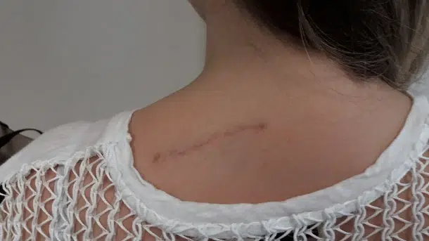
Advantages
- The first and accessory rib removal is easy and safe since there are no overlaying nerves and vessels.
- The brachial plexus is shallow posteriorly, allowing the surgeon to decompress it more effectively Additionally, subclavian vessels are deeper into the brachial plexus and do not interfere with manipulation. Therefore, decompression is easy, safe, and effective.
- The published clinical results concern neurogenic TOS. Whether the same approach suits arterial or venous TOS is decided case by case.
- The rib can be removed up to its joints, so no rib is left behind. This does not rule out recurrence from scar tissue or other causes.
- Nerves and vessels are handled less during bone removal, which is intended to lower the risk of nerve and vessel injury.
- Dr. Aghayev has performed this procedure since 2015; his 2023 paper reports 80 operations in 61 patients.6 All patients improved. Some had complications (an opened pleura, Horner syndrome or a bleed at the top of the lung), and none was permanent. Two patients had already been operated on at other centres, by the anterior or transaxillary approach; they improved too. It is a retrospective series from one surgeon; it does not compare PURED with other approaches.
References
- Nuutinen H, Kärkkäinen JM, Mäkinen K, et al. Long-term outcome over a decade after first rib resection for neurogenic thoracic outlet syndrome. Interact Cardiovasc Thorac Surg. 2021;33(5): 734-740. https://doi.org/10.1093/icvts/ivab172[↩]
- Jammeh ML, Yang A, Abuirqeba AA, et al. Reoperative Brachial Plexus Neurolysis After Previous Anatomically Complete Supraclavicular Decompression for Neurogenic Thoracic Outlet Syndrome: A 10-Year Single-Center Case Series. Oper Neurosurg (Hagerstown). 2022;23(2): 125-132. https://doi.org/10.1227/ons.0000000000000252[↩]
- Clagett OT. Research and prosearch. J Thorac Cardiovasc Surg. 1962;44:153-166[↩]
- Aghayev K, Ciklatekerlio O. Posterior Upper Rib Excision for Neurogenic Thoracic Outlet Syndrome—Feasibility and Early Outcomes. Oper Neurosurg (Hagerstown). 2018;14(5): 532-537. https://doi.org/10.1093/ons/opx143[↩]
- Akaslan I, Ertas A, Uzel M, Ozdol C, Aghayev K. Surgical Anatomy of the Posterior Intermuscular Approach to the Brachial Plexus. Hand (N Y). 2021;16(6): 759-764. https://doi.org/10.1177/1558944719895619[↩]
- Aghayev K. Safety and Efficacy of Posterior Upper Rib Excision and Decompression Technique for Surgical Treatment of Neurogenic Thoracic Outlet Syndrome. World Neurosurg. 2023;180: e739-e748. https://doi.org/10.1016/j.wneu.2023.10.017[↩]



